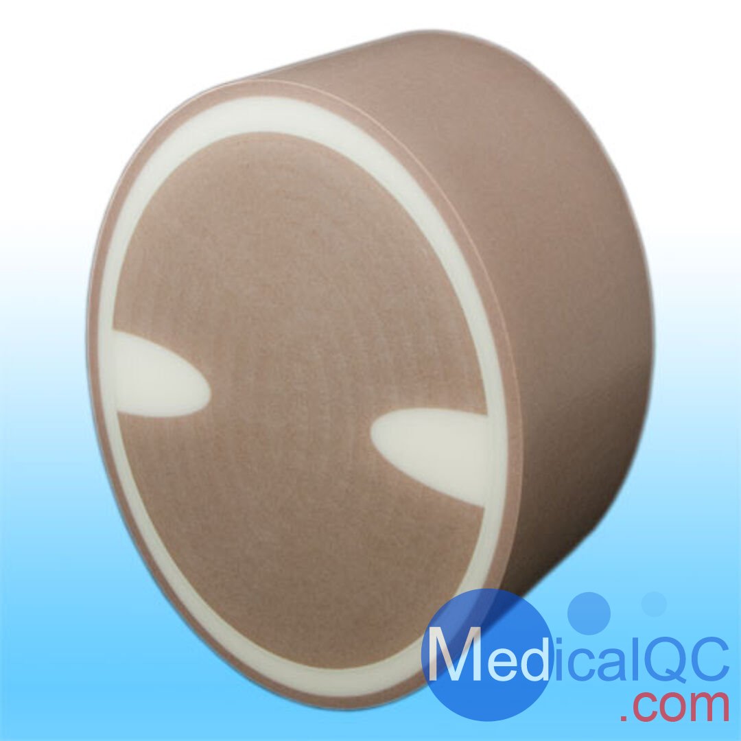
QRM-Cranial-CT-Phantom,颅脑CT模型,Cranial CT Phantom
型号:QRM-Cranial-CT-Phantom 类别: 质控模体 品牌:德国QRM pdf资料: QRM-Cranial-CT-Phantom,颅脑CT模型,Cranial CT Phantom.pdf
QRM-Cranial-CT-Phantom,颅脑CT模型,Cranial CT Phantom用于 CT/FD-CT 成像的人脑解剖结构模拟
QRM-Cranial-CT-Phantom,颅脑CT模型,Cranial CT Phantom基于具有不同 kV 设置的恒定 CT 值的软组织等效塑料和具有类似于人类头骨的 X 射线吸收的周围骨骼结构,人类大脑的两个不同部分可用。
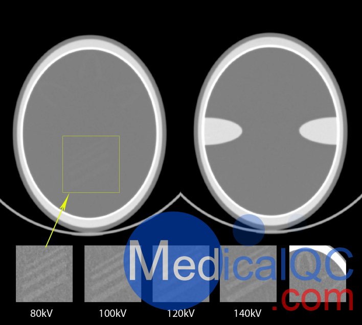
一节描述了具有与颞骨相同的均匀软组织和骨结构的颅底。本部分适用于演示和评估颅底的图像质量,特别是受光束硬化效应、部分体积效应和扫描仪不稳定引起的伪影(例如阳极摆动)的影响。
第二部分旨在测试成像系统的低对比度能力。在均匀的软组织内,几个低对比度结构模拟了大脑的典型皮层结构和中央低对比度结构。


QRM-Cranial-CT-Phantom,颅脑CT模型,Cranial CT Phantom技术参数:
尺寸:180 毫米 x 140 毫米 x 85 毫米组织材料:
密集的骨头: 1 毫米
中等致密骨: 8 / 4 毫米
皮肤(组织): 4 毫米
软组织: 标称 35 HU (80 t 140 kVp, ± 5 HU)
低对比度结构: + 7 HU (±2 HU) 高于周围组织
中等致密骨: 400 毫克 HA/立方厘米
密集的骨头: 1000 毫克 HA/立方厘米

The cranial phantom mimics the
human brain anatomy for CT imaging.
Based on a soft tissue equivalent plastic with a constant CT-number for the different kV settings and a surrounding bone structure with an X-ray absorption similar to the human skull, two different section of the human brain are realized.
One section describes the base of the skull with uniform soft tissue and bony structures equivalent to the temporal bones. This section is suitable to demonstrate and evaluate the image quality in the base of the skull, especially affected by beam hardening effects, partial volume effects and artifacts caused by scanner instabilities, e.g. anode wobble.
The second section was designed to test the low contrast capabilities of the imaging system. Within the uniform soft tissue several low contrast structure simulating typical cortex structures and central low contrast structures of the brain.
SAG:
QRM-Cranial-CT-Phantom,QRM颅脑CT模体,颅脑CT模体,Cranial CT Phantom,QRM脑模体,QRM颅脑CT模型,颅脑模型