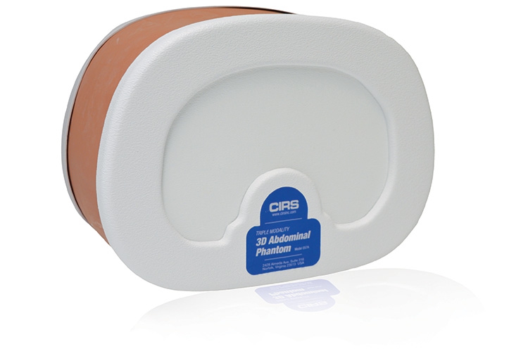
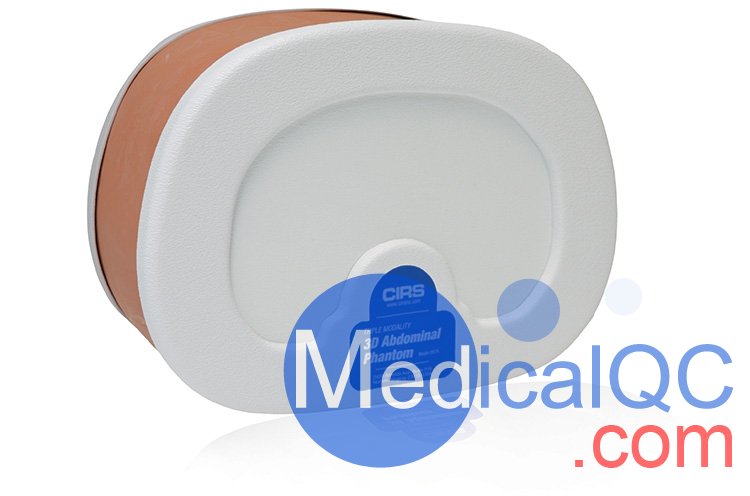
CIRS 057A 3D腹部模体,CIRS 057A腹部模体
型号:CIRS 057A 类别: 质控模体 品牌:美国CIRS pdf资料: CIRS 057A 3D腹部模体,CIRS 057A腹部模体.pdf
CIRS 057A 3D腹部模体,CIRS 057A腹部模体
CT /超声波/ MRI图像融合 - 实时扫描 - 活组织检查培训
CIRS 057A 3D腹部模体
CIRS 057A 3D腹部模体采用Zerdine®(1)的自我修复配方,允许以最少的针迹跟踪进行多次活检插入,是演示影像导航技术的理想选择。
腹部成像对于诊断疾病和监测治疗是有用的。057A型是小型成人腹部的代表,可在CT,MR和超声下进行成像。此功能使幻像成为图像融合研究等应用的有用工具; 成像协议的发展; 扫描技术培训; 和系统测试,验证和演示。
CIRS 057A 3D腹部模体使用简化的拟人几何模型将腹部从大约胸部椎骨(T9 / T10)模拟到腰椎(L2 / L3)。这些材料提供了在CT,MR和超声波下的结构之间的对比。穿刺时,固体聚合物背景凝胶不会泄漏。*
CIRS 057A 3D腹部模体内部结构包括肝脏,门静脉,两个部分肾脏,部分肺,腹主动脉,腔静脉,模拟脊柱和六肋骨。肝脏有六个病灶,每个肾脏有一个病灶。肌肉层和外部脂肪层包围这些结构和塑料端盖使幻像足够持久的扩展扫描。
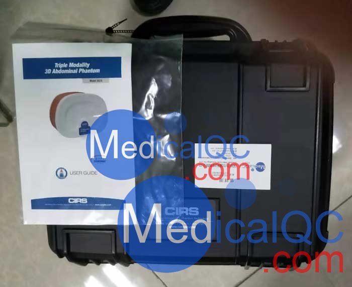
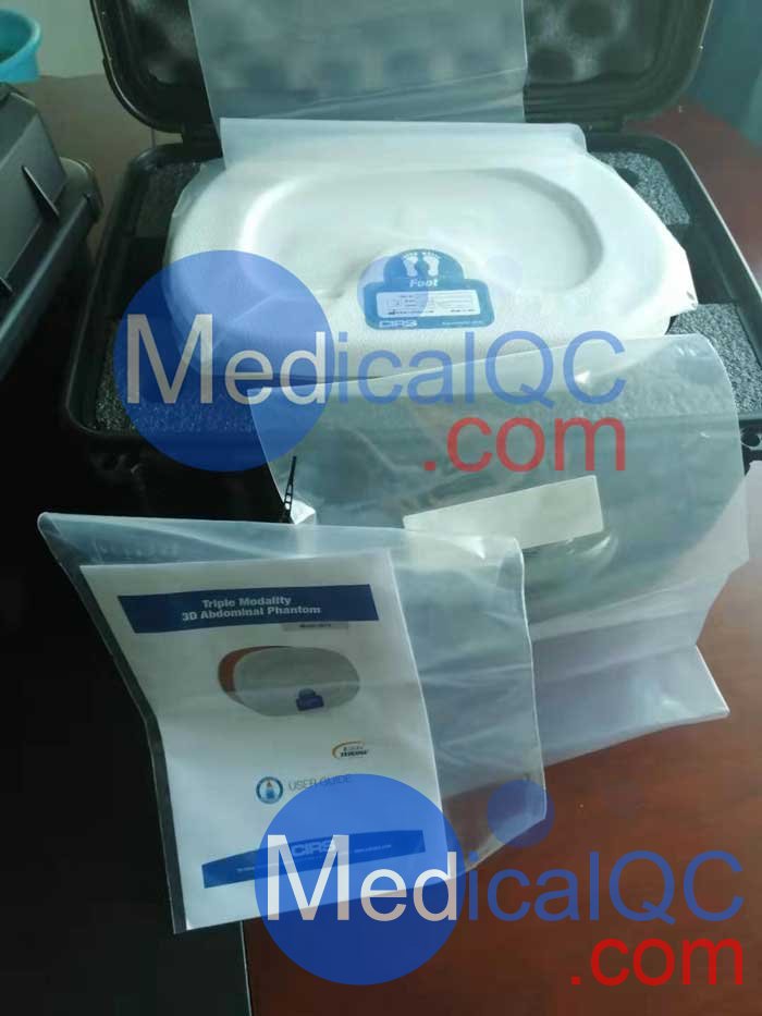
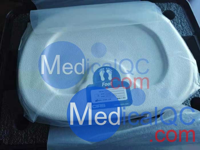
CIRS 057A 3D腹部模体包括泡沫衬里硬手提箱。为了适应图像融合技术,CIRS可以提供增值选项和服务,如幻像专用CMM,参考CT或MRI数据集,客户特定注册设备的附件以及包含特殊点标记。
CIRS 057A 3D腹部模体特征:
演示CT,超声波和MRI扫描技术
评估图像融合算法
测试新设备
验证自动活检系统
优化成像协议
改善徒手腹部活检的表现
CT/ ULTRASOUND/ MRI IMAGE FUSION• LIVE SCANNING• BIOPSY TRAINING
The CIRS Triple Modality 3D Abdominal Phantom is constructed of a self-healing formulation of Zerdine®(1) that allows multiple biopsy insertions with minimal needle tracking, and is ideal for demonstrating image-guided navigation technologies.
Abdominal imaging is useful for diagnosing disease and monitor-ing treatments. The Model 057A is representative of a small adult abdomen and can be imaged under CT, MR and ultrasound. This feature makes the phantom a useful tool for applications such as image fusion studies; imaging protocol developments; scan technique training; and system testing, validation and demonstration.
The Model 057A simulates the abdomen from approximately the thorax vertebrae (T9/T10) to the lumbar vertebrae (L2/L3) using simplified anthropomorphic geometry. The materials provide con-trast between the structures under CT, MR and ultrasound. The solid polymer background gel will not leak when punctured.*
Internal structures include the liver, the portal vein, two partial kidneys, a partial lung, the abdominal aorta, the vena cava, a simulated spine and six ribs. The liver has six lesions and the kidneys each have one lesion. A muscle layer and outside fat layer surround these structures and plastic end caps make the phantom durable enough for extended scanning. Blood vessels
have CT contrast added to provide enhanced auto registration in image fusion applications
The phantom includes a foam lined hard carry case. For users interested in image fusion studies, the phantom can be pur-chased as a kit to include a serial-number specific CT DICOM Data set for reference. CIRS can also offer value-added options and services such as phantom specific CMM, reference CT
or MRI data sets, attachment of customer specific registration devices and inclusion of special point markers.
Features
• Demonstrate CT, ultrasound and MRI scan techniques
• Assess image-fusion algorithms
• Test new equipment
• Optimize imaging protocols
• Improve performance of freehand abdominal biopsies
CT /超声波/ MRI图像融合 - 实时扫描 - 活组织检查培训
CIRS 057A 3D腹部模体
CIRS 057A 3D腹部模体采用Zerdine®(1)的自我修复配方,允许以最少的针迹跟踪进行多次活检插入,是演示影像导航技术的理想选择。
腹部成像对于诊断疾病和监测治疗是有用的。057A型是小型成人腹部的代表,可在CT,MR和超声下进行成像。此功能使幻像成为图像融合研究等应用的有用工具; 成像协议的发展; 扫描技术培训; 和系统测试,验证和演示。
CIRS 057A 3D腹部模体使用简化的拟人几何模型将腹部从大约胸部椎骨(T9 / T10)模拟到腰椎(L2 / L3)。这些材料提供了在CT,MR和超声波下的结构之间的对比。穿刺时,固体聚合物背景凝胶不会泄漏。*
CIRS 057A 3D腹部模体内部结构包括肝脏,门静脉,两个部分肾脏,部分肺,腹主动脉,腔静脉,模拟脊柱和六肋骨。肝脏有六个病灶,每个肾脏有一个病灶。肌肉层和外部脂肪层包围这些结构和塑料端盖使幻像足够持久的扩展扫描。
CIRS 057A 3D腹部模体包括泡沫衬里硬手提箱。为了适应图像融合技术,CIRS可以提供增值选项和服务,如幻像专用CMM,参考CT或MRI数据集,客户特定注册设备的附件以及包含特殊点标记。
CIRS 057A 3D腹部模体特征:
演示CT,超声波和MRI扫描技术
评估图像融合算法
测试新设备
验证自动活检系统
优化成像协议
改善徒手腹部活检的表现

| 产品规格 | |
| 外形尺寸 | 26厘米×12.5厘米×19厘米(10.2“×4.9”×7.5“) |
| 幻影重量 | 11磅 (5公斤) |
| 物料 |
外壳:ABS 外脂肪层:Z-皮肤™ 硬组织:环氧树脂 肺:聚氨酯 等软组织:Zerdine ®凝胶 |
| 内部器官 |
肝脏6个病变(2各自小,中,大) (2)肾脏(1个介质病变各) (1)脊柱 (1)偏肺 (1)门静脉 (1)腔静脉 (1)主动脉 (6)的肋 周围软组织(2大病变) |
| 型号057A包括 |
三重模态3D腹部幻影 用户指南 6个月保修 |
| 可选功能 | 型号035: CT DICOM数据组(特定序列号,120 kpv的切片厚度为1.5 mm) |


CT/ ULTRASOUND/ MRI IMAGE FUSION• LIVE SCANNING• BIOPSY TRAINING
The CIRS Triple Modality 3D Abdominal Phantom is constructed of a self-healing formulation of Zerdine®(1) that allows multiple biopsy insertions with minimal needle tracking, and is ideal for demonstrating image-guided navigation technologies.
Abdominal imaging is useful for diagnosing disease and monitor-ing treatments. The Model 057A is representative of a small adult abdomen and can be imaged under CT, MR and ultrasound. This feature makes the phantom a useful tool for applications such as image fusion studies; imaging protocol developments; scan technique training; and system testing, validation and demonstration.
The Model 057A simulates the abdomen from approximately the thorax vertebrae (T9/T10) to the lumbar vertebrae (L2/L3) using simplified anthropomorphic geometry. The materials provide con-trast between the structures under CT, MR and ultrasound. The solid polymer background gel will not leak when punctured.*
Internal structures include the liver, the portal vein, two partial kidneys, a partial lung, the abdominal aorta, the vena cava, a simulated spine and six ribs. The liver has six lesions and the kidneys each have one lesion. A muscle layer and outside fat layer surround these structures and plastic end caps make the phantom durable enough for extended scanning. Blood vessels
have CT contrast added to provide enhanced auto registration in image fusion applications
The phantom includes a foam lined hard carry case. For users interested in image fusion studies, the phantom can be pur-chased as a kit to include a serial-number specific CT DICOM Data set for reference. CIRS can also offer value-added options and services such as phantom specific CMM, reference CT
or MRI data sets, attachment of customer specific registration devices and inclusion of special point markers.
Features
• Demonstrate CT, ultrasound and MRI scan techniques
• Assess image-fusion algorithms
• Test new equipment
• Optimize imaging protocols
• Improve performance of freehand abdominal biopsies
SAG:
CIRS 057A,CIRS 057A腹部模体,057A腹部模体,CIRS 057A腹部模体,CIRS 057A模体